Figure 6.
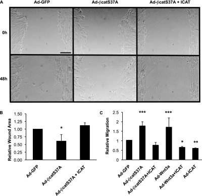
Wnt/β-catenin signaling stimulates fibroblast migration. (A) NHLFs were “scratch” wounded, infected with the stated adenoviruses, and photographed at 0-, 24-, and 48-hour time points. Representative images for each infection and 48-hour time point are shown. (B) Quantification of wound size at 48 hours. Results are expressed as mean change ± SD of wound area relative to wound area at 0 time point from four independent experiments (*P < 0.05). (C) Lung fibroblasts (NL-57) obtained from a patient without pulmonary disease were infected with Ad-GFP, Ad-S37A–β-catenin, Ad-ICAT, and Ad-Wnt3a and analyzed in a Boyden chamber assay as described in Materials and Methods. Similar results were observed with NHLFs (Figure E3). Results are expressed as the mean number of cells migrated in each condition relative to control adenovirus ± SD from eight independent experiments (*P < 0.05; **P < 0.01; ***P < 0.001).

