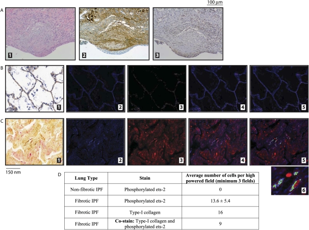
Figure 4.
Lung sections from patients with idiopathic pulmonary fibrosis (IPF) exhibit increased concentrations of phosphorylated ets-2 that colocalizes with Type I collagen. (A) Lung sections from patients with IPF were obtained from the storage bank at the Department of Pathology of Ohio State University Medical Center. Patients were selected according to their clinical diagnosis of interstitial lung disease and IPF. Embedded slides were first stained with H&E (part 1) to verify the pathological appearance of fibroblastic foci, indicative of usual interstitial pneumonia. Serial sections were then stained with Type I collagen (part 2) and phosphorylated ets-2 (part 3). (B and C) Lung tissues were obtained and classified as described in Materials and Methods. Nonfibrotic areas of IPF lungs (B) and fibrotic areas of IPF lungs (C) were stained for Type I collagen (brown signal) and phosphorylated ets-2 (red signal) (part 1). The Nuance imaging system then converted the Type I collagen signal to blue (part 2) and the phosphorylated ets-2 signal to red (part 3). The merging of parts 2 and 3 is shown in part 4. The Nuance imaging system then overlaid the images and converted those cells that stained positive for both Type I collagen and phosphorylated ets-2 to green (part 5). A magnification of the colocalization of Type I collagen and phosphorylated ets-2 in part 5 is shown in part 6. (D) Quantitation of images from B and C.

