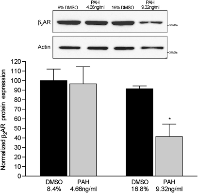
Figure 2.
Concentrations of membrane-bound β2–adrenergic receptors (β2ARs) in murine tracheal epithelial cells exposed to PAHs for 24 hours. Western blot represents whole-cell membrane fractions probed with an anti-human β2AR antibody. Graphical data represent optical density of a 52-kD band normalized to same sample actin concentrations to control for interlane loading variations, and then to control cells exposed to vehicle only (8.4% and 16.8% DMSO in complete medium). Band intensity was arbitrarily set at 100 for control cells treated with 8.4% DMSO, and normalized to actin. *P < 0.01, versus all other groups.

