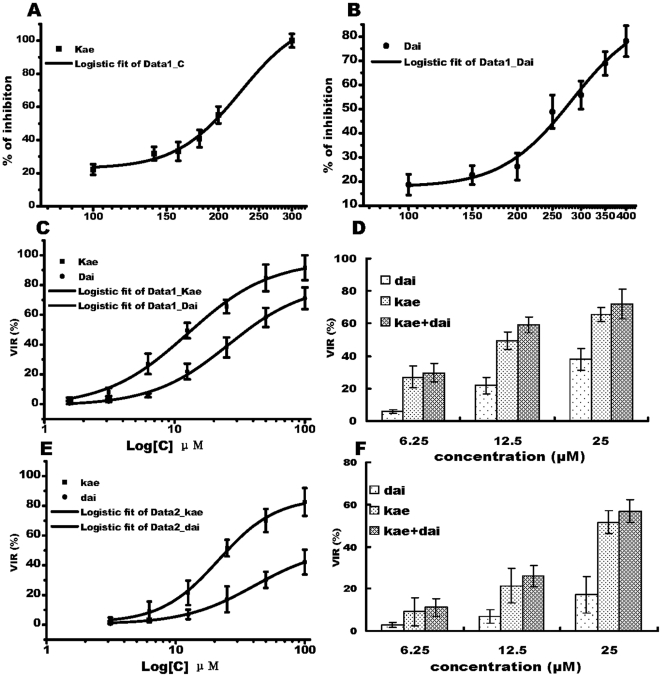
Figure 1. Toxicity of Kae and Dai on BHK21 cells and their effects on JEV infection.
(A) Growth inhibition curve of Kae on BHK21 cells for 72 h. CC50 was 230 µM. (B) Growth inhibition curve of Dai on BHK21 cells for 72 h. CC50 was 270 µM. (C) Anti-JEV effects of Kae and Dai when cells were pretreated for 2 h before infection with 0.1 MOI JEV for 72 h. EC50 for Kae and Dai at 72 h were 12.6 and 25.9 µM, respectively. (D) Comparison of anti-JEV effects of combination treatment with Kae and Dai when cells received combined pretreatment for 2 h, and were then infected with 0.1 MOI JEV for 72 h. (E) Anti-JEV effects of Kae and Dai when cells were infected with 0.1 MOI JEV for 2 h and then treated with Kae or Dai for 72 h. EC50 was 21.5 µM for Kae and 40.4 µM for Dai. (F) Comparison of anti-JEV effects of combination treatment with Kae and Dai when cells were infected with 0.1 MOI JEV for 2 h followed by combination treatment for 72 h. The data represent the means for five replicate samples of three separate experiments. Kae (black square) or Dai (black circle).

