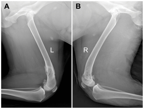
Figure 2. Radiological analysis of intracorporeally and in situ devitalized bone segment.
(A) Plain radiograph showed periosteal callus formation covering the entire cortical surface of the devitalized bone segment at 3 months. (B) Partial sclerosis still remained within the microwave-treated bone segment at 12 months.

