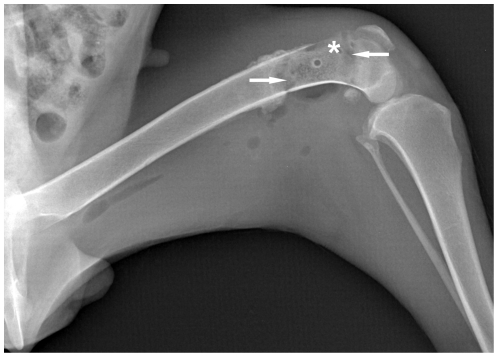
Figure 3. Radiological manifestation of bone resorption.
Taken at 2 months after surgery, this radiograph showed a variant trend towards bone resorption. In this radiograph, the bone mineral density of the targeted bone segment was decreased, and in particular two crescent-shaped radiolucent lesions (arrows) were developed in its two ends between the dead bone segment and the normal bone tissues. In addition, focal cortical bone in the targeted bone segment became thinner or disappeared due to resorption (asterisk).

