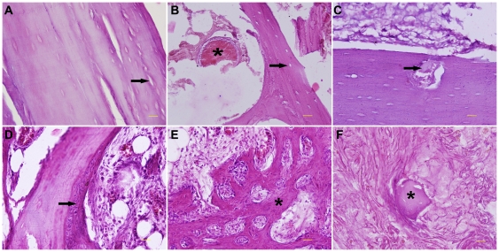
Figure 6. Histological analysis about the healing process of intracorporeally devitalized bone segment.
(A–F) Haematoxylin and eosin staining. (A) Necrotic cortex with empty osseous lacunae (arrow) at 2 weeks. (B) Coagulative necrotic tissues (asterisk) and acellular trabeculae (arrow) in bone marrow cavity at 2 weeks. (C) Initial infiltration of fibrovascular tissues (arrow) into the dead bone at 2 months after surgery. (D) Partial substitution of dead trabeculae by new woven bone (arrow) at 3 months. (E) Newly formed trabecuale (asterisk) at 9 months. (F) Remnants of dead trabeculae (asterisk) surrounded by amounts of fibrovascular tissues at 12 months. Scale bars: 100 µm.

