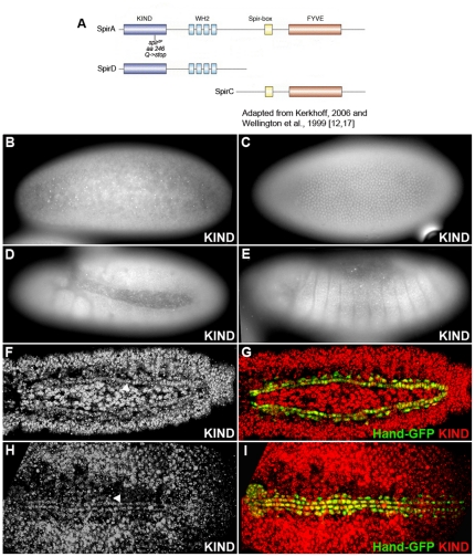
Figure 1. Spir expression pattern during Drosophila embryogenesis.
(A) Diagram of SpirA, SpirC and SpirD proteins and the location of spir2F mutant. Anti-KIND antibody recognizes KIND domain in both SpirA and SpirD proteins. Anti-SpirD antibody recognizes SpirD and the N-terminus of the A-isoform. (B–I) Whole-mount antibody stainings of wild-type embryos with anti-KIND. Anti-SpirD antibody exhibits same staining pattern (data not shown). (B) Stage 4. (C) Stage 5. Focus on the lateral epidermis. (D) Stage 11. Fully extended germ band. (E) Stage 13 (lateral view). (F, G) Stage 15. Dorsal view to visualize the distinct accumulation of Spir in cardioblasts and pericardial cells. (H, I) Stage 16 (Dorsal view). Hand-GFP is expressed in cardioblasts, pericardial cells and the lymph gland. The Spir expression is co-localized with Hand-GFP in heart cells. Arrowheads point to cardioblasts.

