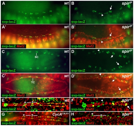
Figure 3. Pattern of Tin- and Svp-positive cells in wt, CycAC8LR1 and spir2F embryos.
(A, A′, B and B′) Lateral view at stage 13. (C, C′, D and D′) Dorsal view at stage 15. (E–H) Dorsal view at stage 16. Mef2 (red) stains Mef2-positive cardioblasts and β-Gal (green) stains Svp-positive cardioblasts. In spir2F mutants, when two nuclei appear together, most cells express only Svp. When three or four nuclei appear together, they are stained with both anti- β-Gal and anti-Mef2. SC, Svp-positive cardioblasts; TC, Tin-positive cardioblasts. Arrows point to Svp-positive cardioblasts; Arrowheads point to Svp-positive pericardial cells. * indicates Svp-positive cells; — indicates Tin-positive cells.

