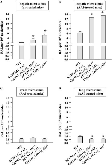
FIG. 4.
DNA adduct formation by AAI in microsomes isolated from CYP1A-humanized and WT mice (Arlt et al., 2011a), untreated (A) or pretreated with AAI (B–D). (A) Hepatic microsomes from untreated mice were used. Hepatic (B), renal (C), and pulmonary (D) microsomes from mice pretreated with AAI were used. Values are given as means ± SD of three parallel incubations (n = 3). RAL, relative adduct labeling. Comparison was performed by t-test analysis; *p < 0.01, different from WT.

