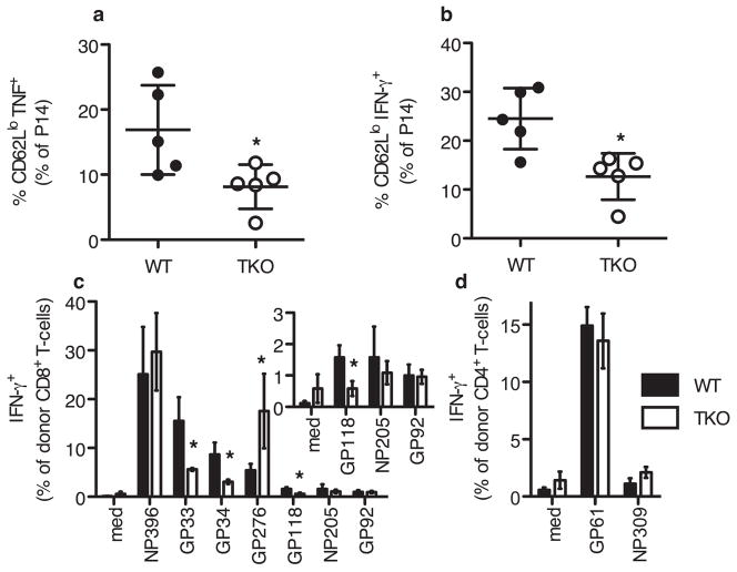
Fig. 4. Altered T cell responses in LCMV-infected TKO mice.
(a,b)Two days post LCMV infection, WT and TKO animals were injected with BFA and BFA-treated P14 Tg splenocytes. Spleens were harvested 4 hours later, stained (surface and intracellular) and analyzed by flow cytometry. Representative of 3 experiments. (c,d)To prevent rejection of adoptively transferred cells, host T cells were depleted in WT Thy1.1+Thy1.2+ and TKO Thy1.1+Thy1.2+ mice using anti-Thy1.2. 3×10⁷ LCMV-immune WT Thy1.1 splenocytes were injected i.v. one day before LCMV infection. 5.5 days post-infection, animals were sacrificed and spleens were harvested. Cells analyzed by in vitro restimulation followed by surface and intracellular cytokine staining. Mean and standard deviation of 3 animals of each strain, representative of two experiments. Inset shows low abundance CD8 T-cell responses, enlarged from (c). Asterisk indicated P<0.05, by two-tailed, unpaired t-test.

