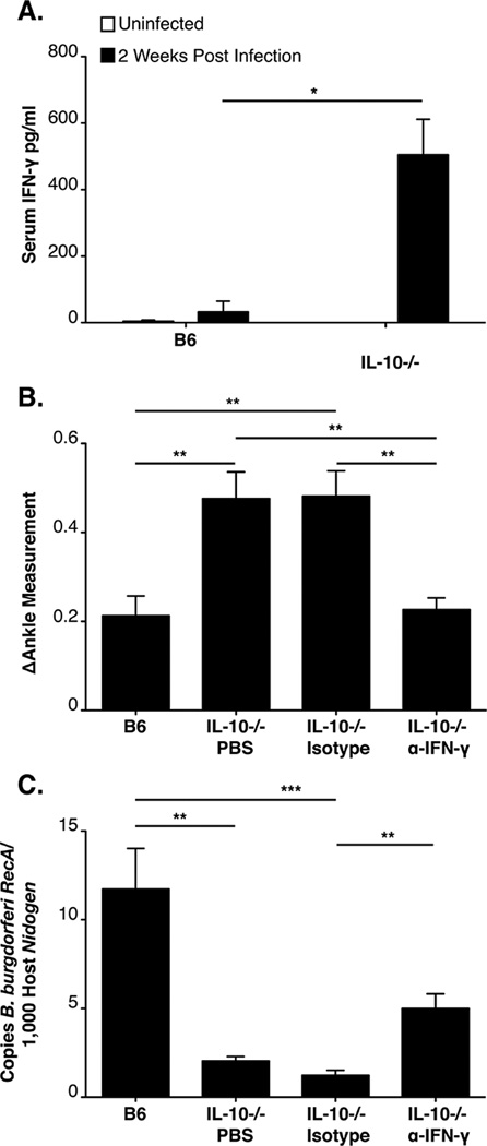
Figure 4. IFN-γ is responsible for the increased arthritis severity in B6 IL-10−/− mice.
ELISA of serum IFN-γ at 2 weeks of infection in B6 and B6 IL-10−/− mice (N=5 mice per group (A). Arthritis was assessed in B6 and B6 IL-10−/− mice at 4 weeks of infection (N≥8 mice per group). B6 IL-10−/− mice were injected with PBS, control antibody, or α-IFN-γ over the course of infection. Ankle swelling was used to assess arthritis severity (B). Quantification of joint spirochetes at 4 weeks of infection, using qPCR (C). Statistical analyses were performed as reported in Materials and Methods.

