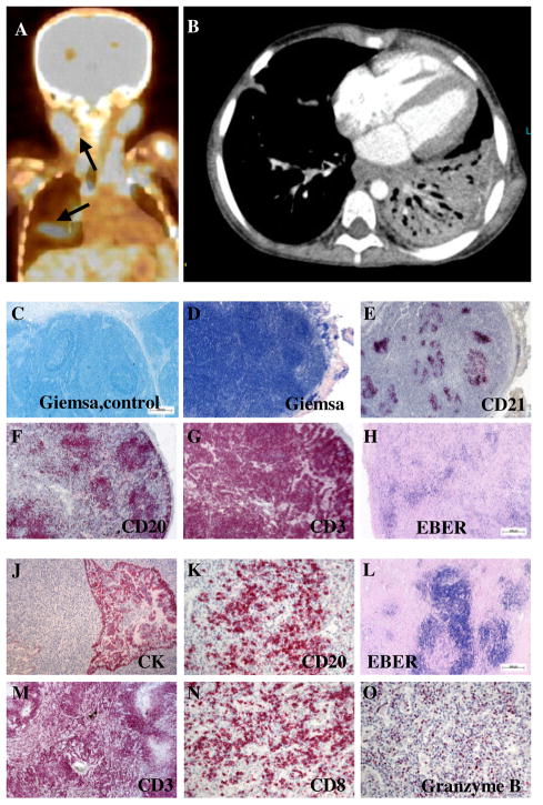
Figure 2. Destructive EBV positive lymphoproliferative disease.
A, Positron emission tomography (PET) scan of P5. Arrows indicate areas of high metabolic activity reflecting lymphoproliferative disease. B, Pulmonary CT scan of P5 showing a dense infiltrate of the left lower lobe. C-H, Lymph node histology: Giemsa staining of a tumor-free lymph node from a control patient with cancer (C) and the lymph node from P5 (D). Immunohistological staining of follicular dendritic cells (anti-CD21; E), B cells (anti-CD20; F) and T cells (anti-CD3; G). The anti-EBER (H) staining shows the presence of EBV infected cells. J-O, Lung histology: Immunohistological staining of cytokeratin (J), B cells (anti-CD20; K) and T cells (anti-CD3; M), that were predominantly CTL (anti-CD8; N) expressing high amounts of Granzyme B (O). The lung lesions were also highly positive for EBV infected cells (anti-EBER; L). The magnification was 2,5x (C-F,H and L), 5x (G,J and M) and 20x (K,N and O).

