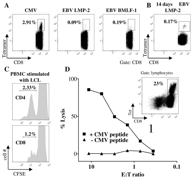
Figure 5. Antiviral T cells are generated and respond to antigen stimulation in vitro.
A, Frequency of CD8+ T cells binding HLA-A2 tetramers labelled with epitopes of CMV (pp65) and EBV (LMP-2 and BMLF-1). B, Staining of CD8+ T cells with LMP-2 tetramer after 14 days culture of PBMC from the patient with autologous irradiated PBMC pulsed with LMP-2 peptide. C, CFSE dilution of CD4+ (upper panel) and CD8+ T cells (lower panel) incubated with autologous EBV-LCL for 3 days. D, PBMC were cultured with CMV pp65 peptide for 3 weeks leading to an expansion of virus-specific CTL (insert indicates the fraction of CD8+ T cells expressing a pp65 specific TCR as detected by tetramer staining). Lytic activity of these cells was tested on autologous 51Cr loaded EBV-LCL target cells that were used unpulsed (triangles) or after incubation with CMV p65 peptide (squares). Spontaneous lysis was below 30%.

