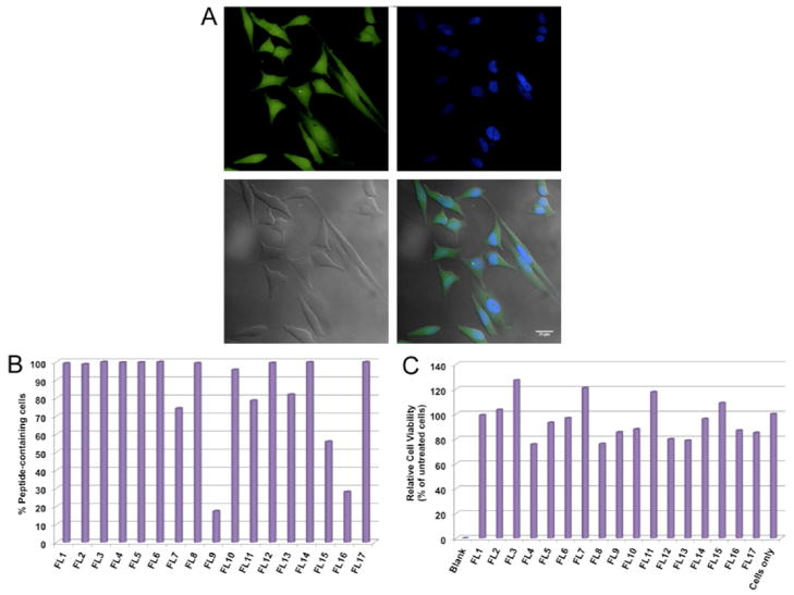
Figure 4.
A) Cellular uptake of branched peptides into HeLa cells, top left: fluorescence image of cells; top right: DAPI staining of the nucleus; bottom left: phase contrast image; bottom right: overlay of all images. White scale bar is 25μm. B) Cell permeability of FITC-labeled branched peptides by flow cytometry in HeLa cells. C). MTT cell toxicity assay.

