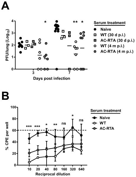
Figure 3. Contribution of antibody to AC-RTA-mediated protection.
A, The number of PFU per lung 3 or 7 d after WT γHV68 challenge in mice treated with sera from naïve mice, mice 30 d or 4 m after WT infection, or mice 30 d or 4 m after AC-RTA vaccination. *P≤0.05, **P≤0.01, one way ANOVA. B, The percent of wells positive for cytopathic effect (CPE) after in vitro infection with WT γHV68 pre-treated with naïve serum, or sera from mice 4 m after WT infection or AC-RTA vaccination (n=2-5/group). Dotted line is at 60.3%, the average percent of CPE when no serum was added. ns, not significant, *P≤0.05, **P≤0.01, ***P≤0.001, one way ANOVA.

