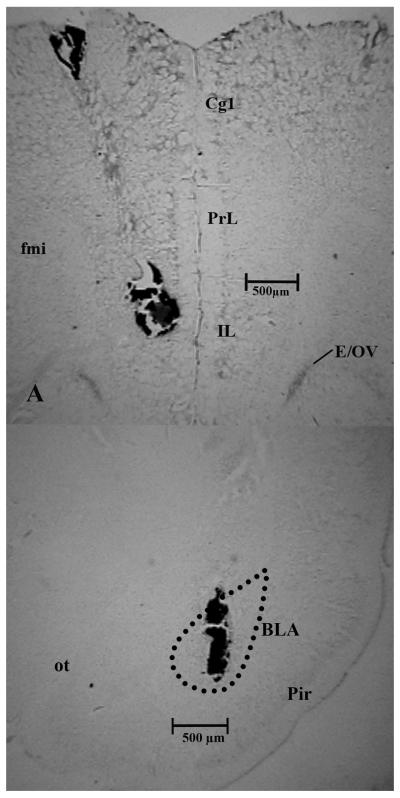
Figure 2a and 2b.
Representative photomicrograph is shown of a coronal brain section of the mPFC (2a) and BLA (2b). The needle tract and ink injection are clearly visible and indicate an injection site approximately 3.2 mm (mPFC) and 0.2 mm (BLA) rostral to bregma. Injection volume of the ink was identical to the drug/vehicle volume used in the experiments. Abbreviations: mPFC – medial prefrontal cortex, Cg1 – cingulate cortex, area 1, PrL – prelimbic cortex, IL – infralimbic cortex, E/OV – ependymal layer/olfactory ventricle, fmi – forceps minor corpus callosum, BLA – basolateral amygdala, Pir – piriform cortex, ot – optic tract.

