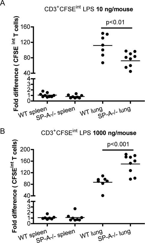
Figure 1. SP-A can either enhance or suppresses exogenous stimulation induced T cell activation in the lung in vivo.
Mice were oropharyngeally instilled with low (10 ng/mouse) or high (1000 ng/mouse) LPS in conjunction with CFSE. Single cell suspensions from spleen and lung digests were prepared after 68-72 h, stained with T cell markers and analyzed for CFSE-int (divided cells) by flow cytometry (1A-B). Data is normalized to WT spleen, and represented as the fold difference in CFSE-int T cells from either 8 or 9 mice per group, with three independent experiments. Proliferating BrdU+ T cells in lungs or spleen were identified by intracellular flow cytometric staining in conjunction with surface markers. * p<0.05 compared to WT mice.

