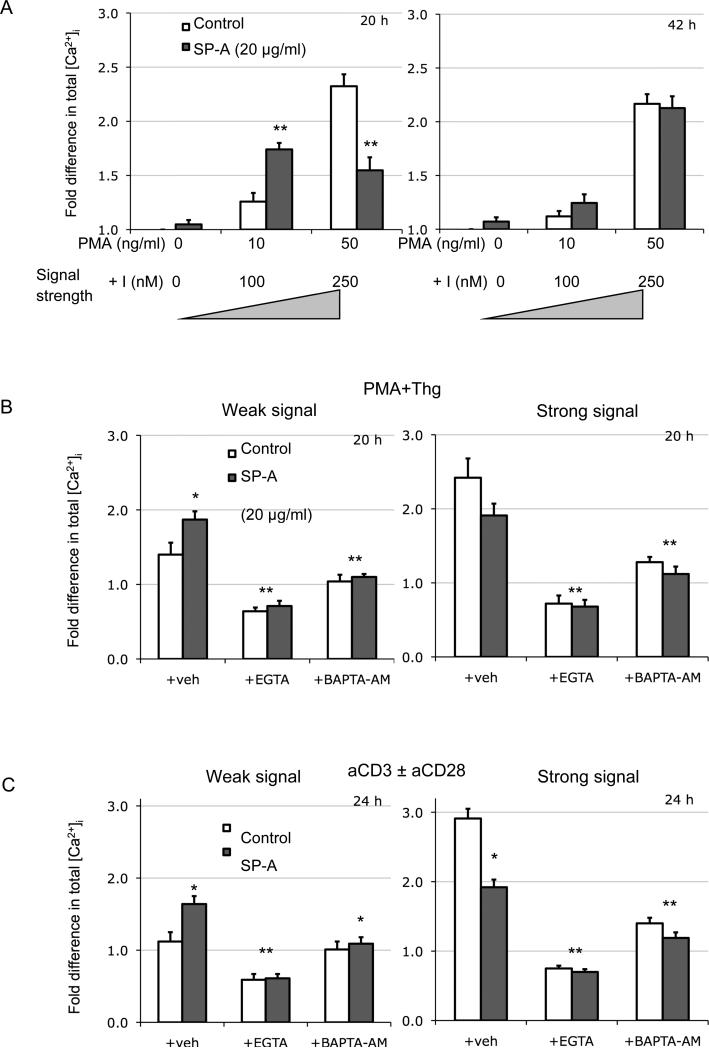
Figure 6. [Ca2+]i levels are enhanced in the presence of SP-A at lower signal strengths irrespective of mode of T cell activation.
[Ca2+]i measurements were performed from cells activated for various periods with low or high doses of PMA+I (7A), PMA+Thg (7B) or anti-CD3 ± anti-CD28 (7C) in the absence or presence of SP-A (20 μg/ml). During the last 60 mins of activation, cells were also pulsed with Fluo-4 and analyzed by flow cytometry. Two distinct time points representing the two [Ca2+]i profiles are depicted in Figure 7A. In 7B and C, cells were also suspended in buffer containing EGTA or BAPTA-AM during activation. All data is normalized to baseline [Ca2+]i levels in cells alone conditions at the indicated time points post-activation, and are representative of 3 or 4 independent experiments with pooled cells from the lungs of 5 mice. *p<0.05, **p<0.01 compared to respective control condition.

