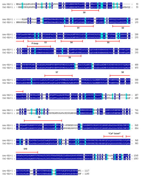
Figure 2.
SLO-1 A. suum and C. elegans and alignments of deduced amino-acid sequences. Identical amino-acids between C. elegans and A. suum sequences are shaded in dark blue and distinct aminoacids sharing similar physico-chemical properties are shaded in light blue. Predicted signal peptide sequences are shaded in grey. The transmembrane domains (TM) are noted below the sequences. Comparison of Asu-SLO-1 (Genbank accession n° ACC68842.1) and Cel-SLO-1 (Genbank accession n° NP_001024259.1) showing the 7 TM domains (S0 – S6), P-loop, S7 – S10 intracellular domains and “Ca2+ bowl”. Domain annotation corresponds to A. suum SLO-1. Figure, modified from Buxton et al., 2011.

