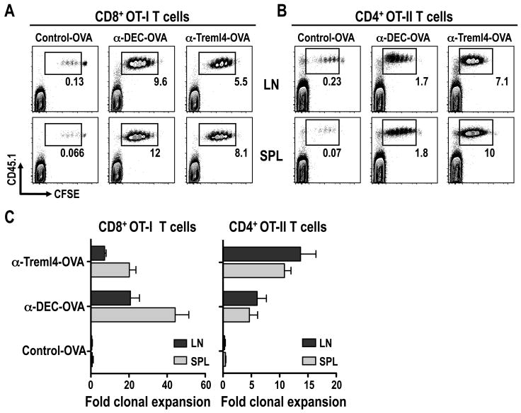
Figure 4. α-Treml4-OVA induces in vivo proliferation of CD8+ and CD4+ transgenic T cells.
B6 mice were injected i.v. with 2-5 × 10⁶ CFSE-labeled OT-I (A) or OT-II (B) T cells, and 24 hrs later with 3 μg Control-, α-DEC- and α-Treml4 -OVA fusion mAbs s.c. footpad. Three days after mAb inoculation, skin draining LN (LN) and spleen (SPL) were harvested and expansion of CD8+ or CD4+ Vα2+ T cells was evaluated by FACS for CFSE dilution. Shown is the percentage of transferred CD45.1+ T cells undergoing more than one division. Plots are gated in CD4+ CD3ε+ T cells, and are representative of three experiments. (C) As in A-B, but bar graph quantified T cells undergoing more than one division (fold clonal expansion; see materials and methods) as the mean +/- SD of 4-6 animals in 3-4 different experiments.

