
Figure 4.
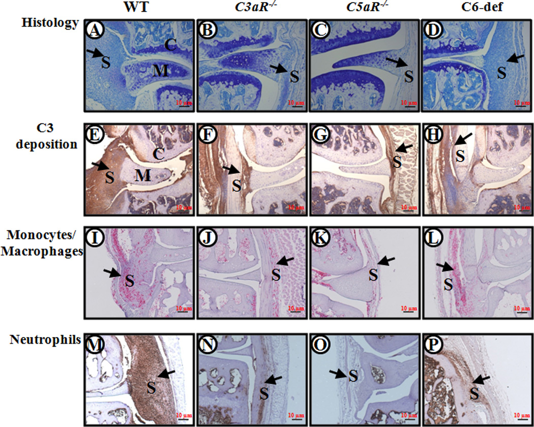
Histopathology, C3 deposition, monocyte/macrophage infiltration, and neutrophil infiltration from WT, C3aR−/−, C5aR−/−, and C6-def mice with CAIA. Knee joints with maximum CDA scores from WT, C3aR−/−, C5aR−/−, and C6-def mice with CAIA were selected. Areas of synovium (S), cartilage (C), and meniscus (M) are identified. The black arrows all point to the synovium. From left to right, knee joints of WT, C3aR−/−, C5aR−/−, and C6-def mice stained with Toluidine-blue (blue color) are shown in A, B, C and D. Similarly, knee joints with C3 deposition (brown color) are shown in E, F, G and H; knee joints stained with F4/80 (red color) for monocytes/macrophages are shown in I, J, K and L; and knee joints stained with neutrophil (brown color) surface marker are shown in M, N, O and P. Magnification for all pictures was 10X (a magnification red scale bar at 10X of 10 µm (0.01mm) is included on the lower right hand corner in all frames).