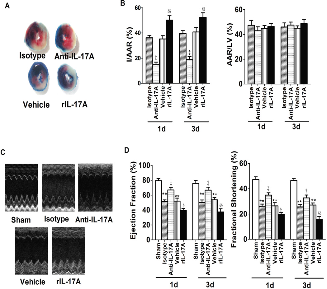
Fig. 1.
IL17A neutralization and repletion affected mouse myocardial I/R injury. A, Representative images of LV slices from different groups at 1 day after reperfusion. The nonischemic area is indicated in blue, the AAR in red and the infarct area in white. B, Quantification of infarct size of myocardial tissues 1 day or 3 days after reperfusion (n=6–8). C, Representative M-mode echo-cardiography images of the left ventricular 1 day after reperfusion. D, Ejection fraction and LV fractional shortening (n=8). **P<0.01 versus sham; †P<0.05, ‡P<0.01 versus isotype; §P<0.05, §§P<0.01 versus vehicle.

