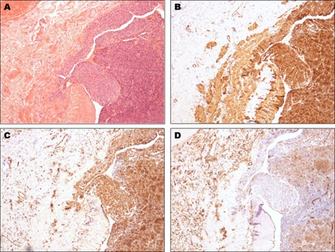
Figure 2.
(A) Figure 1 at x100 magnification. (B) Smooth muscle actin staining both the glomus cells and the smooth muscle of the vessel walls (original magnification x100). (C) Strong vimentin staining of the glomus cells (original magnification x100). (D) Patchy CD34 staining of the glomus cells with background staining of endothelial cells and interstitial dermal dendritic cells (original magnification x100).

