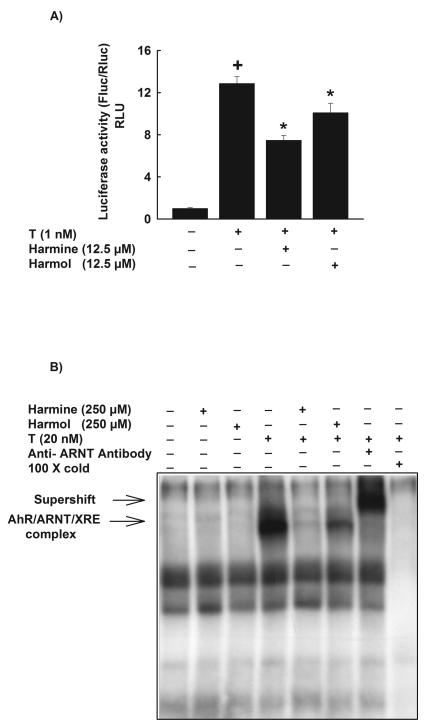
Figure 5. Effect of harmine and harmol on XRE-luciferase activity and AhR activation using electrophoretic mobility shift assay (EMSA).
A, HepG2 cells were transiently co-transfected with XRE-luciferase reporter plasmid pGudLuc 6.1. and renilla luciferase control plasmid pRL-CMV. Cells were treated with DMSO, harmine (12.5 μM) or harmol (12.5 μM) 30 min before the addition of TCDD (1nM) for an additional 24 h. Cells were lysed and luciferase activity is reported as relative light unit (RLU) of firefly luciferase to renilla luciferase (Fluc/Rluc) (mean ± S.E.M., n = 4). (+) P < 0.05 compared with Control (C), (*) P < 0.05 compared with TCDD (T). B, In vitro AhR activity was measured by EMSA using guinea pig hepatic cytosolic extracts. Cytosolic extracts were incubated with DMSO, harmine (250 μM) or harmol (250 μM) for 30 min before the addition of TCDD (20 nM) for 2 h. The mixtures were tested for binding activity to a [γ-32P]-labeled XRE consensus oligonucleotide for an additional 15 min. The products of this binding were separated on a 4% polyacrylamide gel. The specificity of the binding was confirmed by competition assays using anti-ARNT antibody or a 100 fold molar excess of unlabeled XRE. AhR-ARNT-XRE complex formed on the gel was visualized by autoradiography. One representative of three experiments is shown.

