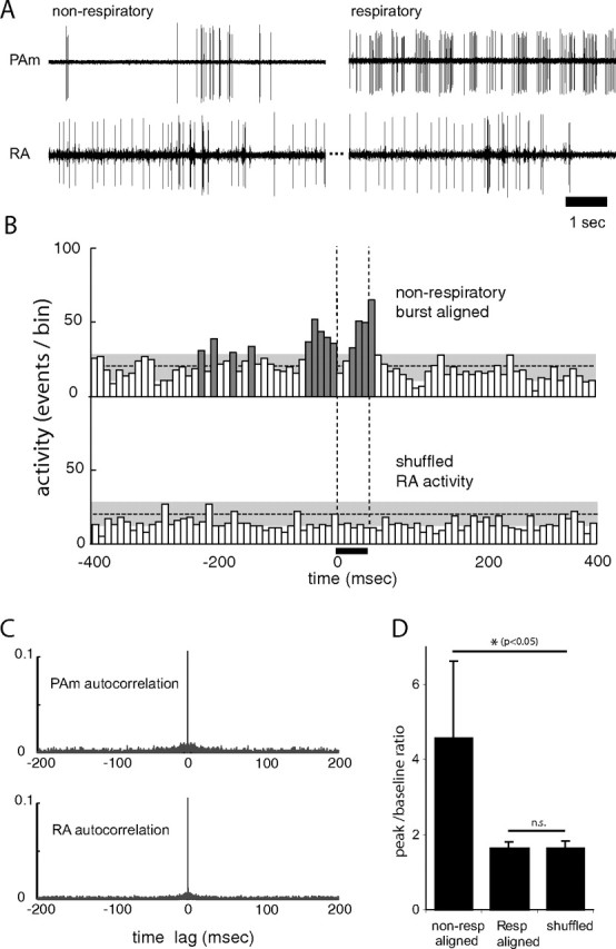
Figure 7.

Spontaneous activity of nonrespiratory neurons in PAm is correlated with bursting in the contralateral RA. A, Simultaneous recordings of two single units in the left PAm while maintaining the same unit in the contralateral RA. For the left pair, the recording electrode was placed closer to a site showing the characteristic nonrespiratory bursting pattern. Bursts in these neurons occur at approximately the same time as bursts (and subsequent pauses) in RA. In the right pair, a respiratory neuron recorded several minutes after the unit on the left shows a complete lack of correlated activity with the same RA neuron. B, Two PETHs are shown, representing activity recorded in the right RA of one bird. In the top PETH, RA activity was aligned to spikes from the nonrespiratory PAm neuron. In the bottom PETH, RA activity was randomly selected from the same recording. Significant peaks in the top histogram (dark gray) indicate that RA activity is high around the time of bursts in the PAm nonrespiratory bursting neurons, but shows no correlation with the shuffled control. The horizontal dotted lines indicate the mean height of histogram peaks generated from randomly selected 50 ms periods of RA activity. The gray areas indicate two SDs from that mean. The vertical dotted lines delineate the window of time for all PETHs used to calculate the values shown in D. C, Autocorrelation plots of PAm (top) and RA (bottom) activity used in B, showing values up to 0.1 (of 1.0). The absence of peaks at time lags greater or smaller than 0 indicate that the activity seen in the PETHs does not result from intrinsic correlation in RA or PAm. D, Comparison of peak/baseline ratios for RA activity 50 ms after alignment (0–50 ms) to nonrespiratory bursting neurons (left), and respiratory neurons (middle), and when the nonrespiratory aligned RA activity is randomly shuffled (right). Error bars indicate SD. Asterisks indicate significance by paired t test.
