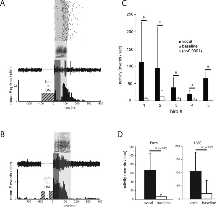
Figure 8.
Nonrespiratory PAm neurons are active during vocalization evoked under anesthesia. A, An example single-unit recording made from PAm. The raster at the top shows spike times (black) from one PAm neuron overlaid on periods of vocal production (gray) for 40 instances of DM stimulation-evoked vocalizations. The sonogram beneath the raster represents a sample vocalization. The neural trace below represents sample activity from the PAm neuron during that vocalization. The light gray box surrounding the sample indicates the vocalization period for this example. The PSTH below the trace was compiled from the 40 trials. To examine only vocalization-related activity and exclude activity resulting from direct trans-synaptic activation, the first 50 ms of activity after stimulation (dark gray box) was ignored when performing the analysis shown in C and D. B, A multiunit example from another bird. A longer period of stimulation was used (600 ms) than in A. Unlike in A, activity at this site was elevated at an earlier time point relative to vocalization, indicating a heterogeneous temporal relationship between PAm nonrespiratory neurons and EVOC production. C, Mean ± SD of neural activity in PAm recorded during multiple instances of DM stimulation evoked vocalizations (black) and paired baseline periods (immediately before each stimulation, white) for five birds. D, Population activity (grand mean ± SD) recorded during evoked vocalizations for PAm (left) and HVC (right). PAm activity was gathered from five sites in four birds, and HVC activity was gathered from four sites in three birds. Asterisks indicate significance by paired t test.

