
Figure 2.
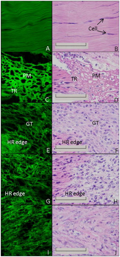
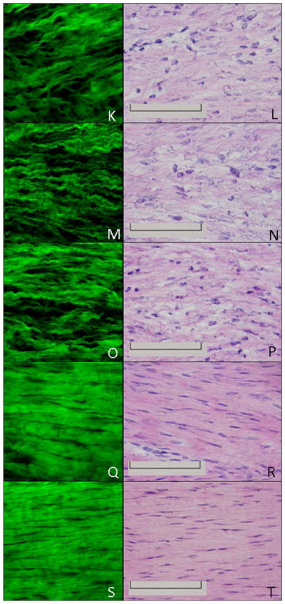
H&E and MPLSM of the healing ligament. MPLSM staining (left column) and the corresponding H&E image (right column) of the normal intact (A-B) or healing ligament at 1 day (C-D), 3 days (E-F), 5 days (G-H), 7 days (I-J), 9 days (K-L), 11 days (M-N), 14 days (O-P), 21 days (Q-R) or 28days (S-T) post injury. TR: transected region; PM: provisional matrix; GT: granulation tissue; HR edge: healing region edge; Original Magnification: 600X