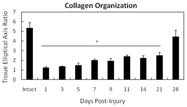
Figure 3.
Quantification of collagen disorganization using the TEAR metric (TEAR=1 represents randomly oriented fibers. Higher values show greater organization). Normal ligament (CX) exhibits greater collagen organization indicated by a higher TEAR. Injury significantly (p < .05) reduces collagen organization from days 1-21. Collagen orientation of the day 28 healing ligament is similar to the uninjured MCL (p=0.056). Asterisk indicates significance differences between the intact ligament and the day 1-21 healing ligaments. Significance was based on p < .05.

