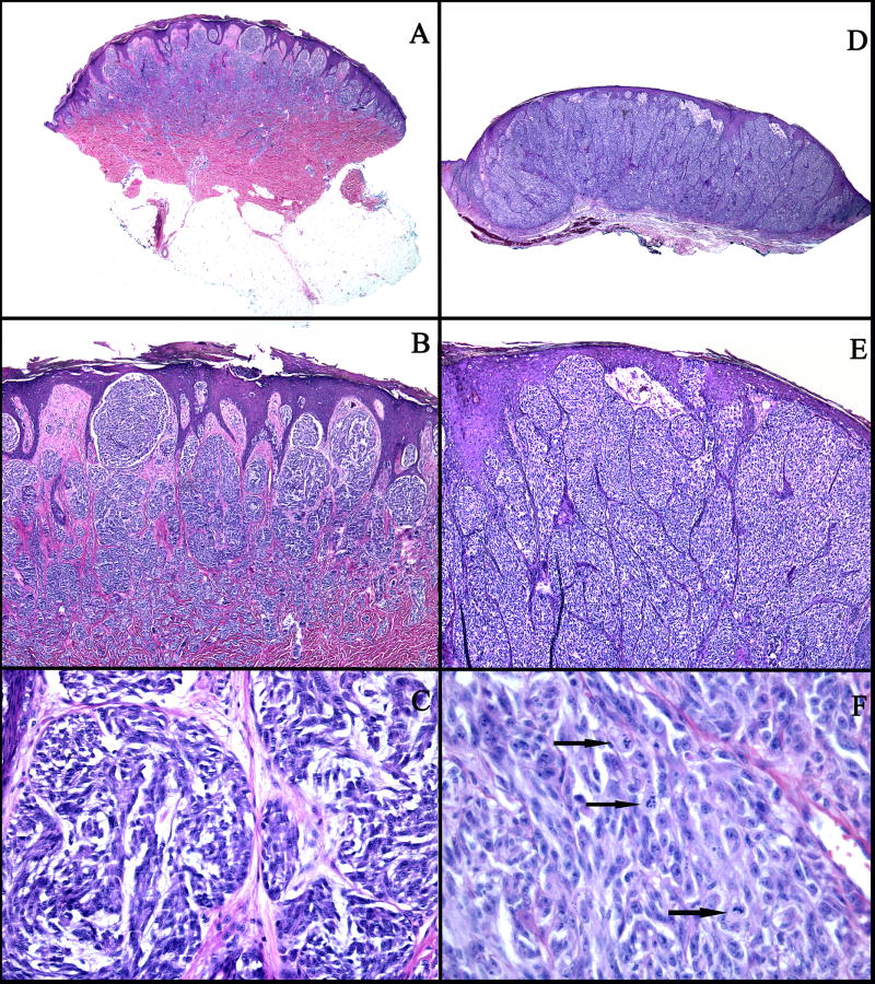
Figure 1.
A–C: An example of a compound SN (case 1): A: Low power view showing a symmetric, well-circumscribed, and wedge-shaped proliferation of melanocytes in the epidermis and in the dermis. B: There are large vertical nests of melanocytes with clefts between the nests and the surrounding hyperplastic epidermis. C: The melanocytes are large, slightly pleomorphic, with vesicular nuclei, prominent nucleoli, and abundant pale cytoplasm. D–F: An example of a SMM (case 33): D: Asymmetric and poorly circumscribed proliferation of melanocytes in the epidermis and dermis. E: Large confluent nests of melanocytes, focally forming sheets in the dermis, and irregular nests and single melanocytes in the epidermis. F: Markedly pleomorphic melanocytes with vesicular nuclei, prominent nucleoli, eosinophilic cytoplasm, and numerous mitotic figures (arrows).

