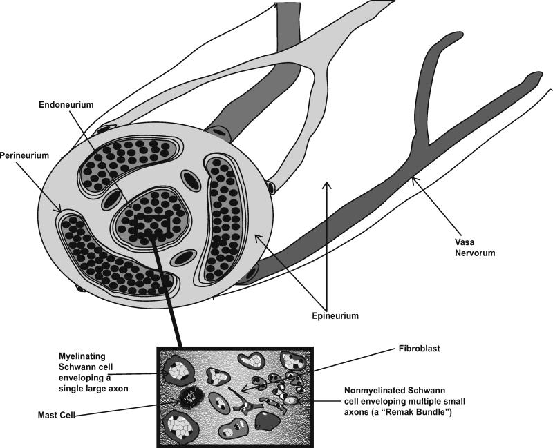
Fig. 1. Schematic illustrating the anatomy of normal peripheral nerve.
Indicated are the outmost layer of nerve (the epineurium) and the vasa nervorum, the perineurium (which ensheathes bundles of nerve fibers and forms the blood-nerve barrier) and the endoneurium. The inset highlights the mixture of cell types present in the endoneurium including axons, myelinating and nonmyelinating Schwann cells, fibroblasts and mast cells.

