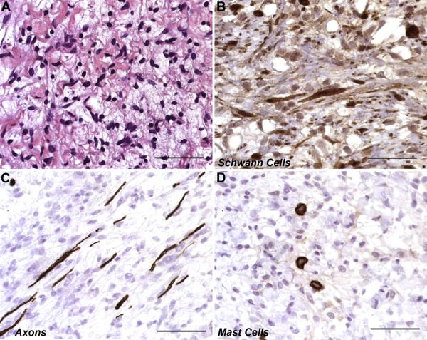
Fig. 2. Photomicrographs demonstrating the presence of multiple cell types in neurofibromas.
Unlike other types of peripheral nerve sheath tumors, neurofibromas are composed of a complex mixture of multiple cell types normally present in peripheral nerve. (A) Hematoxylin and eosin stained section of a plexiform neurofibroma showing the typical loosely packed collection of spindled cells characteristic of these tumors. (B) Immunostains for S100β label the cytoplasm and nuclei of Schwann cell-like elements within a plexiform neurofibroma. (C) Axons, which are visualized here by their immunoreactivity for neurofilaments, are separated by neoplastic Schwann cells and other cellular elements recruited into the tumor. This demonstrates the infiltrative nature of these lesions. (D) Numerous mast cells, identifiable by their immunoreactivity for CD117 (c-Kit), are also present in this plexiform neurofibroma. Scale bars, 50 μm.

