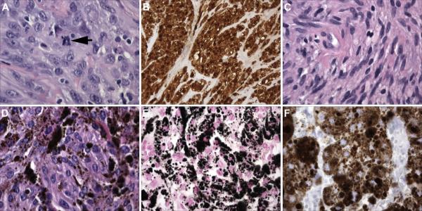
Fig. 3. A comparison of the pathology of MPNSTs with that of conventional schwannomas and melanotic schwannomas, two other tumor types composed predominantly of neoplastic Schwann cells.
(A) Hematoxylin and eosin stained section of an MPNST showing the high degree of cellularity and nuclear atypia characteristic of these neoplasms. The arrow indicates a mitotic figure. (B) Unlike plexiform neurofibromas, the benign precursors from which they arise, MPNSTs are overwhelmingly composed of neoplastic cells which in this panel are highlighted by their S100β immunoreactivity. (C) In contrast, this schwannoma resected from the VIIIth cranial nerve of an NF2 patient shows a lower degree of cellularity and relatively uniform “cigar-shaped” tumor cell nuclei. (D) Hematoxylin and eosin stained section of a melanotic schwannoma, a tumor type associated with Carney complex. Note the abundant deposits of brown pigment. (E) A Fontana stain highlights the melanin in a melanotic schwannoma as black deposits. (F) Unlike conventional schwannomas, melanotic schwannomas express antigens characteristic of melanocytes and melanomas. Shown is an immunostain for the melanoma marker HMB45 in this melanotic schwannoma. Magnification: A, C, D, E and F; 40x; B, 20x.

