Figure 5.
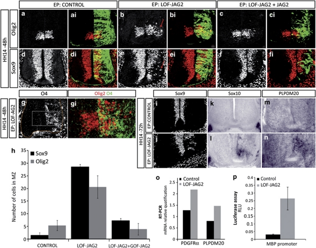
Depletion of Jagged2 activity at the time of MN generation results in premature OLP generation. (a–h) Embryos electroporated at HH14 with the indicated DNAs were analysed at 48 h PE with OLP markers. EP side is shown to the right, GFP (green) shows transfected cells. (a and b) Olig2 is expressed at pMN in control NT, whereas GOF-JAG2 EP shows migratory Olig2+ cells. (c) Co-EP with human Jagged2 (GOF-JAG2) restores Olig2+ cells to pMN. (d and e) Sox9 is expressed throughout the VZ in control NT, whereas LOF-JAG2 electroporated NT shows depletion of Sox9+ cells at pMN and migratory Sox9+ cells. (f) Co-EP with GOF-JAG2 restores Sox9 expression in pMN. (g) Olig2+ migratory cells (red) co-expressed the OLP marker O4 (green). (h) Quantitative analysis of Olig2+ and Sox9+ migratory cells at 48 h PE after electroporation of the indicated DNAs. Numbers are shown as migratory marker expressing cell in each experimental condition. Bars correspond to the standard error (S.E.M.). (i–n) At 72 h PE of LOF-JAG2, Sox9 is extinguished from the VZ, and premature expression of the OLP markers Sox10 and PLPDM 20 are detected by in situ hybridisation. In control electroporated spinal cords, Sox10 and PLPDM20 are only expressed in the ventral roots. (o) Premature OLP generation as revealed by the real-time–PCR detection of a significant increase in the expression of PDGF receptor α and in the myelin-specific gene PLP at 48 h PE of LOF-JAG2. (p) Premature OLP generation as revealed by the increased activity of the MBP-Luc reporter 24 h PE of LOF-JAG2

