Figure 1.
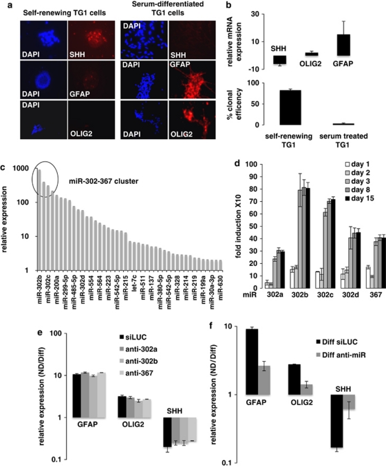
The miR-302-367 cluster contributes to serum-mediated suppression of GiCs stemness. (a) Immunofluorescence and (b, upper panel) QPCR were used to detect SHH and the glial markers GFAP and OLIG2 expression in self-renewing or serum-differentiated TG1 cells. The lower panel of (b) represents the clonal efficiency of self-renewing and serum-treated TG1 cells. (c) Measurement of miRNAs overexpressed after 4 days of serum-mediated differentiation, by Taqman low-density arrayTLDA. The circle highlights the miR-302-367 cluster. (d) QPCR analysis of the miR-302-367 cluster at different time points (1, 2, 3, 8, and 15 days) of serum-mediated differentiation. Self-renewing TG1 cells were cultured either in their defined medium or in 0.5% serum to induce differentiation. At each time point, total RNA was extracted and expression of miR-302a, b, c, d, and 367 was determined using Taqman probe as described in the Materials and Methods section. (e) Anti-miR-302a, anti-miR-302b, anti-miR-367, or (f) a pool of the three anti-miRs were used to transiently transfect TG1 cells to determine the importance of individual or simultaneous functional inhibition of these specific miR in serum-mediated differentiation (Diff). After 4 days of transfection, SHH, GFAP, and OLIG2 expression were assessed by QPCR. siLUC was used as control

