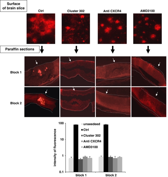
Figure 7.
CXCR4 functional inhibition suppresses TG1 infiltration within a cerebral tissue. TG1 Ctrl cells were pre-incubated or not with the CXCR4 inhibitor AMD3100 or with anti-CXCR4-blocking antibody for 24 h. These cells, as well as TG1 Cluster 302 cells (Cluster 302), were then seeded on the surface of a MBS. The upper panels (surface of brain slice) show cell interactions with the surface of the brain slice. The lower panels (paraffin sections) represent paraffin sections showing cells infiltration only in the control condition (Ctrl). Dashed lines mark the neural tissue boundaries. Block1 and 2 show the section of two different MBSs. The white arrows indicate the surface on which the cells were seeded. The histogram represents a measurement of the fluorescence intensity quantified using the Image J software. The values were compared with the background fluorescence measured in the same conditions in unseeded MBSs

