FIGURE 5.
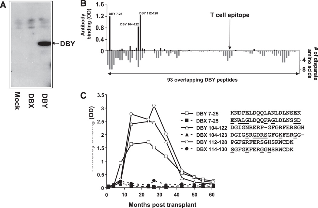
Serologic IgG response to DBY but not DBX after hematopoietic stem-cell transplantation (HSCT). (A). Recombinant DBY and DBX were expressed in human female 293 cells, and total cell lysates were probed in Western blots with plasma collected at 24 months posttransplant. Results with DBY and DBX are compared with 293 cells transfected with an empty vector (mock transfectants). (B). A plasma sample collected from the patient 34 months after transplant was used to map the DBY-specific antibody response. The plasma was diluted 1:50 and tested by ELISA for its reactivity to each of the 93 overlapping DBY peptides (above x-axis). Reactivity to the DBY peptides was revealed with anti-IgG secondary antibodies. The location of the T-cell epitope is indicated by an arrow. The number of disparate amino acids between the DBY and peptides and their DBX counterparts is also represented in this figure (below x-axis). (C). The patient plasma reactivity to the three most immunogenic DBY peptides, 7–25, 104–122 and 112–128, and their DBX homologues was assessed by ELISA using samples collected serially after allogeneic HSCT. Disparate residues between DBX and DBY peptides are represented with underlined characters.

