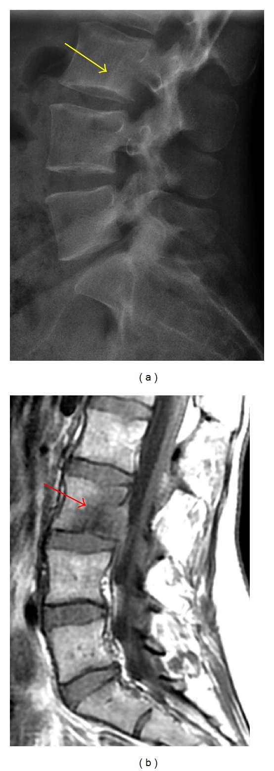
Figure 1.

(a) Lateral radiograph is poor at delineating the L3 vertebral body metastatic lesion, which appears as a faint lucency with a subtle sclerotic margin (yellow arrow). (b) This lesion is better seen on the sagittal T1-weighted MRI as an ill-defined hypointensity (red arrow) within the L3 marrow.
