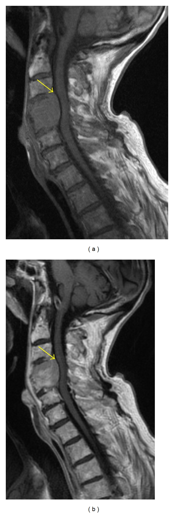
Figure 7.

Sagittal T1-weighted MR image (a) shows a hypointense expansile lesion involving the C4 and C5 vertebral bodies with extension into the ventral epidural space (yellow arrow). The lesion enhances homogeneously on postcontrast T1-weighted MR (b); however, the degree of normal marrow enhancement is similar to that of the metastatic myeloma lesion.
