
Figure 9.
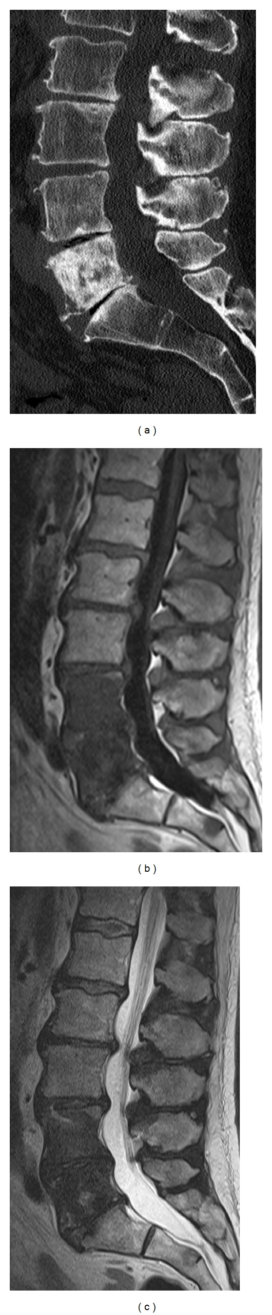
Sagittal CT reformation of the lumbar spines (a) shows a large sclerotic lesion nearly completely involving the L5 vertebral body. Sagittal T1-weighted (b) and T2-weighted (c) images show abnormal hypointense marrow signal in not only the L5 vertebral body but also the L4 vertebral body, corresponding to blastic metastatic lesions.