
Figure 3.
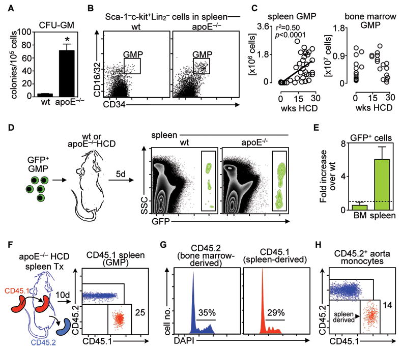
The spleen contains proliferating myeloid cell progenitors that give rise to their progeny in vivo. A. CFU-GM shows colony formation in spleens of wt and apoE−/− HCD mice (means ± SEM, n = 4). *P< 0.05. B. Phenotypic analysis of granulocyte and macrophage progenitors (GMP) in spleens of wt and apoE−/− HCD mice. C, Enumeration of GMP in spleens and bone marrow of apoE−/− mice fed HCD for up to 30 weeks. Linear regression was performed. D. Adoptive transfer of GFP+ GMP to wt and apoE−/− HCD mice. Data show GFP cells in spleens 5 days after transfer. E. Enumeration of data above. Data show the fold increase from wt to apoE−/− HCD mice of adoptively transferred GFP+ cells in the bone marrow (BM) and spleen (data shown are pooled from two independent experiments). F. CD45.1+ spleens from apoE−/− HCD mice were transplanted to asplenic CD45.2+ apoE−/− HCD mice for 10 days. Data show chimerism of GMP in spleens 10 days after transplantation. G. Cell cycle analysis of CD45.1+and CD45.2+ GMP in transplanted spleens shown in G. Numbers indicate percentage of cells in S/G2/M phase (means ± SEM, n = 4). H. Monocyte accumulation in aortic tissue of the mice described in F and G. Representative of 2 independent experiments are shown.