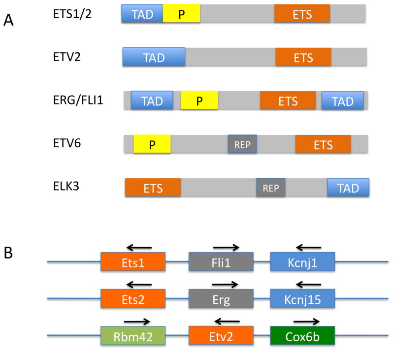
Fig. 1.
ETS proteins expressed in the endothelium. A. Domain structure of ETS factors. The conserved ETS domain (ETS), which includes the sequence specific binding domain, is present in all ETS proteins. The transcription activating domain (TAD) is present in all proteins except ETV6, which only exhibits repressor function. The pointed domain (P) is involved in protein-protein interactions and is present in ETS proteins that can undergo dimerization. ETV6 and ELK3 contain a transcriptional repressor domain (REP). B. Genomic organization Ets1/Ets2/Etv2 subfamily of ETS factors. Note that the Ets1 and Ets2 genes are located adjacent to Fli1 and Erg respectively and that the genomic region shows clear signs of duplication. Although Etv2 is closely related to Ets1 and Ets2 within the ETS domain, the genomic organization surrounding Etv2 is clearly distinct.

