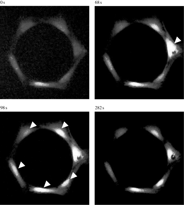
Figure 3.
Fluorescent images in pseudo-colour of a typical intercellular calcium signal propagation process in a looped hexagonal bone cell chain. The cell on the right side has been mechanically stimulated by an AFM indentation probe from 54 s. The [Ca2+]i intensity of the indented cell reached the peak 14 s later. The calcium signal was later transferred to the other cells in the looped chain in both left and right directions. The white arrowheads highlight the cells with peak [Ca2+]i at the image-taken time, which is shown on the top of each image.

