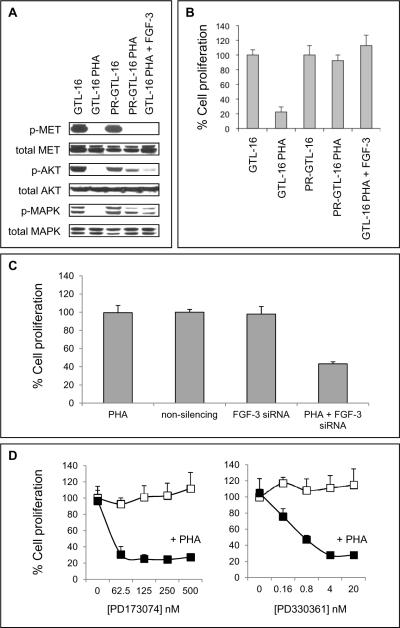
Figure 5. FGF-3 stimulates AKT and MAPK phosphorylation and confers resistance to PHA-665752-mediated growth inhibition.
(A) GTL-16 and PR-GTL-16 cells were treated for 24 h with 400 nM PHA-665752 with or without addition of 0.1 μM recombinant FGF-3, followed by Western Blot analysis. (B) Cells were treated as in (A), followed by a BrdU cell proliferation assay. Data are depicted as mean ± SE. (C) PR-GTL-16 cells were transfected with FGF-3 siRNA and concomitantly treated with 400 nM PHA-665752 for 48 h. Thereafter, cell proliferation was measured using a BrdU cell proliferation assay. For control, cells were transfected with non-silencing siRNA or left untransfected. Data are depicted as mean ± SE. (D) PR-GTL-16 cells were treated with increasing doses of the selective FGF receptor (FGFR) inhibitor PD173074, alone or in combination with 400 nM PHA-665752. In a second set of experiments, PD173074 was replaced by PD330361, another selective small molecule FGFR inhibitor. After 24 h, cell proliferation was measured using a BrdU cell proliferation assay. Open squares = FGFR inhibitor alone, filled squares = FGFR inhibitor in combination with PHA-665752. Data are depicted as mean ± SE.

