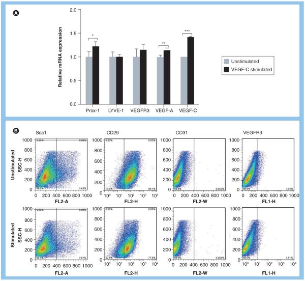
Figure 2. Adipose-derived stem cells stimulated with VEGF-C demonstrate early expression of lymphatic endothelial cell markers and loss of stem cell markers.
Podoplanin staining of tissues harvested from various groups after Matrigel implantation. (A) Relative mRNA expression by adipose-derived stem cells for Prox-1, LYVE-1, VEGF-C, VEGF-A and VEGFR-3 following 48-h exposure to VEGF-C in vitro. Baseline expression of these markers is set to one in unstimulated cells (*p < 0.05, **p < 0.01 and ***p < 0.001). (B) Flow cytometry analysis of isolated adipose-derived stem cells demonstrating expression of stem cell markers (Sca1, CD29) and endothelial and lymphatic endothelial cell markers (CD31, VEGFR3) before (upper panel) and after 48-h stimulation with VEGF-C (lower panel) in vitro.

