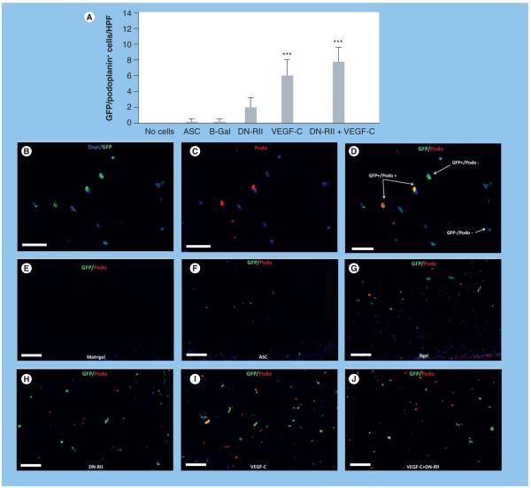
Figure 5. Adipose-derived stem cells express podoplanin in vivo after stimulation with VEGF-C.
(A) Counts of GFP-positive/ podoplanin-positive cells/HPF in Matrigel plugs harvested from various groups (mean + standard deviation; each bar represents sections obtained from three to four HPFs by two independent reviewers per animal with four to five animals per group). Note the significant increase in number of double-positive cells in animals implanted with Matrigel plugs containing ASCs stimulated with VEGF-C as compared with plugs implanted with unstimulated ASCs (***p < 0.001). Transfection with DN-RII caused a modest increase in double-positive cells; however this change was not statistically significant (B-D). Representative florescent double staining of GFP (green; [B]), podoplanin (red; [C]) and overlay ([D] 40× magnification). Dapi stain (blue) was used to stain nuclei of live cells (E-J). Representative double immunofluorescent straining of GFP (green) and podoplanin (red) staining in the various experimental groups (as indicated; 20×). ASC: Adipose-derived stem cell; B-Gal: B-galactosidase; DN-RII: Dominant-negative TGF-β receptor II; GFP: Green fluorescent protein; HPF: High-powered field.

