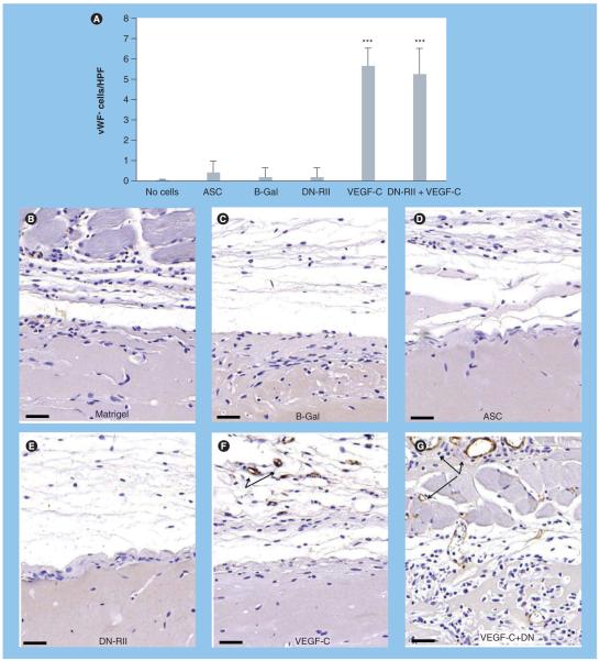
Figure 9. Inhibition of TGF-β signaling does not augment angiogenesis in response to VEGF-C stimulation.
(A) Cell counts of vWF-positive cells/HPF in various experimental groups (mean + standard deviation). Note significant increase in cell counts only in groups stimulated with VEGF-C prior to implantation (***p < 0.001). (B-G) Representative figures (20× magnification) of vWF staining in various experimental groups as noted. Arrows show vWF-positive cells. Scale bars represent 50 μm. ASC: Adipose-derived stem cell; B-Gal: B-galactosidase; DN-RII: Dominant-negative TGF-β receptor II; HPF: High-powered field; vWF: von Willebrand factor.

