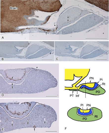
Fig. 1.
Distribution of AQP4 in the rat pituitary gland. Paraffin sections were immunolabeled and visualized using a DAB reaction. Nuclei were stained with hematoxylin. Sagittal sections (A–C) and coronal sections (D, E) are shown. A: Strong AQP4 signals are evident in the infundibulum (arrows) as well as in the brain. This labeling is abruptly diminished in the transitional area between the infundibulum and pars nervosa (arrowhead). The periphery of pars nervosa is also positively stained for AQP4 (double-arrow). B, C: Histochemical controls. Anti-AQP4 antibodies were preabsorbed with an antigen peptide (B) or the antibody was omitted (C). D, E: Examples of average (D) and higher (E) expression of AQP4 from different animals. D: Apparent AQP4 labeling is evident in the periphery of the pars nervosa (arrow), marginal layer (double-arrow), and in the transitional area between the pars distalis and pars intermedia (arrowhead). E: The AQP4 staining pattern is similar to that in D, except for the strong labeling in the region surrounding the cyst-like structures (arrows). F: Schematic drawing of the sagittal section (upper) and coronal section (lower) cut along the line shown in the sagittal section. Inf, infundibulum; PN, pars nervosa; PT, pars tuberalis; PD, pars distalis; PI, pars intermedia; Rt, Rathke’s residual pouch. Bars=1 mm (A–C); 500 µm (D and E).

