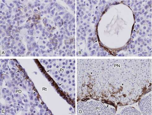
Fig. 2.
Higher magnification views of the localization of AQP4 in the rat pituitary gland. The specimen shown in Figure 1E was examined at a higher magnification. A: AQP4-positive cells are stellate in shape and surround the endocrine cells. B: Cells forming a cyst-like structure are positive for AQP4 (arrows). C: Marginal layer cells covering both pars distalis and pars intermedia are positive for AQP4 (arrows). D: Strong AQP4 signals are localized in the periphery of the pars nervosa (arrow) and in the pars nervosa (arrowheads). PD, pars distalis; PI, pars intermedia; Rt, Rathke’s residual pouch; PN, pars nervosa. Bars=10 µm (A–C); 100 µm (D).

