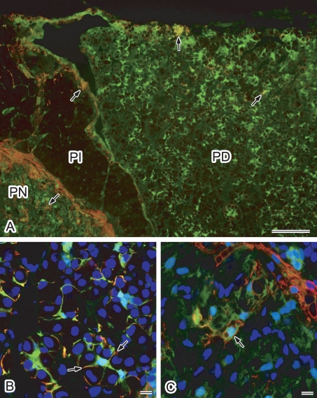
Fig. 3.
Double immunofluorescence localization of AQP4 and S100 protein in the rat pituitary gland. Cryostat sections were labeled for AQP4 (red) and S100 protein (green). Nuclei were stained with DAPI (blue in B and C). A: Conventional fluorescence microscopy at lower magnification. AQP4-positive cells were found to be consistently positive for S100 protein (arrows). Some S100 protein-positive cells were positive for AQP4. B, C: Higher magnification views via laser confocal microscopy. Projection images of consecutive 3-slice confocal images at 1-µm intervals are shown. B: Pars distalis. Labeling for AQP4 accumulates in the processes of S100 protein-positive folliculo-stellate cells (arrows). C: Pars nervosa. Some S100 protein-positive pituicytes are positive for AQP4 (arrow). PD, pars distalis; PI, pars intermedia; PN, pars nervosa. Bars=100 µm (A); 10 µm (B and C).

